Plantar soft tissue stiffness and foot shape
This research was the first to investigate in vivo plantar soft tissue (PST) stiffness in a large sample (N = 100) of healthy adults.
` `

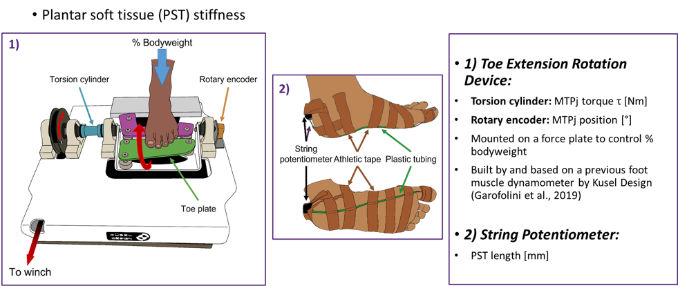
- A manual winch-driven device (custom-built by Kusel Design) was used to extend the metatarsophalangeal (MTP) joint while the foot was subjected to a certain percentage of the participant’s body mass. The load placed on the foot was controlled using a force plate onto which the toe extension rotation device was mounted.
A rotary encoder, attached to one end of the device’s crank shaft, was used to measure the degree of MTP joint extension. Meanwhile, two strain gauges, mounted diametrically onto the torsion cylinder in a full Wheatstone bridge configuration, were used to measure the MTP joint moment. - A string potentiometer threaded through a tube that was attached with adhesive tape to the plantar aspect of the foot along the path of the medial band of the plantar aponeurosis was used to measure PST deformation.
Characteristics of foot shape and foot deformation under load for each participant were determined by 3D scanning and shape and deformation modelling methods described elsewhere.
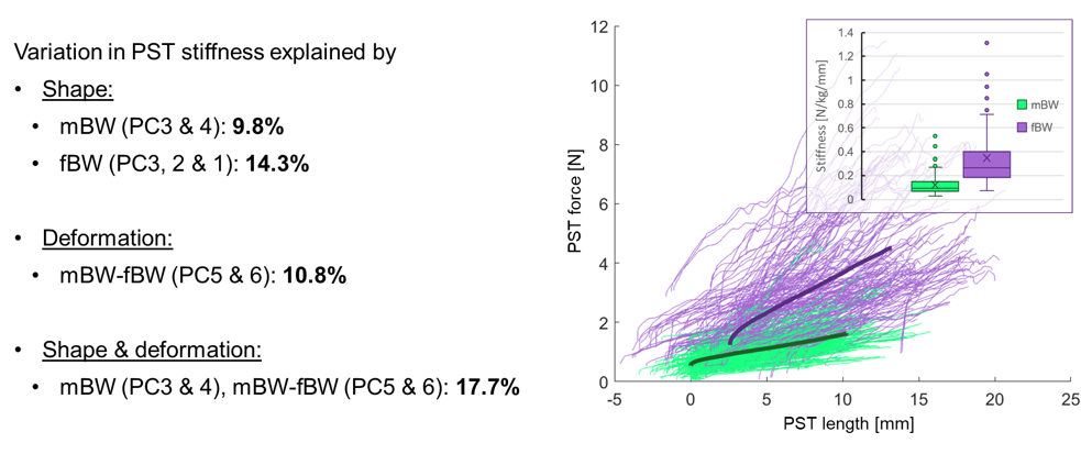
Stepwise multiple linear regression analyses were performed to determine whether variations in foot shape, deformation, as well as their combination, explained variations in PST stiffness.
` `

The results from this research were the first to provide evidence for large variations in PST stiffness across the healthy adult population.
The results also show that variations in foot shape and deformation are poorly related to variations in plantar soft tissue stiffness. Only small proportions of the sample variation in PST stiffness could be explained by characteristics of foot shape, deformation or their combination.
